Mnova BioHOS
Mnova BioHOS
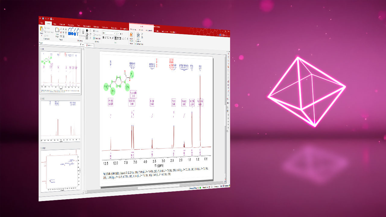
Mnova BioHOS is a specialized NMR data analysis solution for assessing the higher-order structure of biologics or biosimilars such as monoclonal antibodies and other recombinant proteins. It enables objective comparison of test and reference samples using fingerprinting, peak-based metrics, and chemometric approaches applied to 1D and 2D NMR data. By combining advanced spectral processing with statistical modeling, Mnova BioHOS supports robust similarity assessment, classification, and trend detection, helping laboratories ensure structural consistency, product quality, and regulatory confidence throughout development and manufacturing.
Assessment
Features
ECHOS Analysis

CCSD Comparison

1D Profile Analysis

PCA Analysis

PLS Regression

SIMCA-Based Classification

Benefits
Apply Established HOS Strategies: Asses higher-order structure using fingerprint comparison, peak movements, or chemometric analysis against representative reference spectra in one interface.
One Consistent Assessment Framework: Apply fingerprinting, peak-based metrics, and chemometrics under the same preprocessing rules, reducing method-to-method variability.
Reusable Automated Methods: Save, validate and reuse analysis methods to standardize workflows, reduce manual effort and turnaround time.
Reduced Manual Effort and Fewer Transfer Errors: Avoid exporting data to external packages, reformatting matrices, or reprocessing spectra multiple times, which cuts errors and saves time.
Advanced Multivariate Modeling: Analyze complex relationships across multiple variables using proven techniques such as PCA, SIMCA, and PLS for clearer, more reliable insights.
Link Statistics to Spectra with PCA and PLS: Use loadings plots to pinpoint the spectral regions driving dissimilarity and support confident sample classification.
Clear Results Visualization: Review each analysis through contextual plots and linked data tables in a tabbed Workspace, switching between analyses easily without leaving the platform.
Cross-Validation of Conclusions: Confirm findings with complementary approaches, for example ECHOS as a global fingerprint, CCSD for peak movements, PCA or SIMCA for class separation, and Profile for subtle 1D changes.

Publications
- Baldisseri, D., Luo, S., F. Ancajas, C. A., Ortega-Rodriguez, Uriel, Fischer, C., Zou, G., Gu, J., Keire, D., Piotto, M., Zhang, B. NMR-based structural integrity análisis of therapeuctic monoclonal antibodies: a comparative study of Humira and its biosimilars. MAbs. 17, 2551208 (2025). https://doi.org/10.1080/19420862.2025.2551208
- Brinson, R.G., Marino, J.P., Delaglio, F., Arbogast, L.W., Evans, R.M., Kearsley, A., Gingras, G., Ghasriani, H., Aubin, Y., Pierens, G.K., Jia, X., Mobli, M., Grant, H.G., Keizer, D.W., Schweimer, K., Ståhle, J., Widmalm, G., Zartler, E.R., Lawrence, C.W., Reardon, P.N., Cort, J.R., Xu, P., Ni, F., Yanaka, S., Kato, K., Parnham, S.R., Tsao, D., Blomgren, A., Rundlöf, T., Trieloff, N., Schmieder, P., Ross, A., Skidmore, K., Chen. K., Keire, D., Freedberg, D.I., Suter-Stahel, T., Wider, G., Ilc, G., Plavec, J., Bradley, S.A., Baldisseri, D.M., Sforça, M.L., Zeri, A.C.M., Wei, J.Y., Szabo, C.M., Amezcua, C.A., Jordan, J.B., Wikström, M. Enabling adoption of 2D-NMR for the higher order structure assessment of monoclonal antibody therapeutics. MAbs. 11, 94-105 (2019). https://doi.org/10.1080/19420862.2018.1544454
- Hodgson, D. J., Ghasriani, H., Aubin, Y. Assessment of the higher order structure of Humira®, Remicade®, Avastin®, Rituxan®, Herceptin®, and Enbrel® by 2D-NMR fingerprinting. J. Pharm. Biomed. Anal. 163, 144-152 (2019). https://doi.org/10.1016/j.jpba.2018.09.056.
- Emwas, A. H., Saccenti, E., Gao, X., McKay, R. T., Dos Santos, V. A. P. M., Roy, R., & Wishart, D. S. (2018). Recommended strategies for spectral processing and post-processing of 1D 1H-NMR data of biofluids with a particular focus on urine. Metabolomics : Official journal of the Metabolomic Society, 14(3), 31. https://doi.org/10.1007/s11306-018-1321-4
- van den Berg, R. A., Hoefsloot, H. C. J., Westerhuis, J. A., Smilde, A. K., van der Werf, M. J. Centering, scaling, and transformations: improving the biological information content of metabolomics data. BMC Genomics 7, 142 (2006). https://doi.org/10.1186/1471-2164-7-142
- Chen, K., Park, J., Li, F. et al. Chemometric Methods to Quantify 1D and 2D NMR Spectral Differences Among Similar Protein Therapeutics. AAPS PharmSciTech 19, 1011–1019 (2018). https://doi.org/10.1208/s12249-017-0911-1
- Wang, D., Park, J., Patil, S. M., Smith, C. J., Leazer, J. L., Jr., Keire, D. A., Chen, K. An NMR-Based Similarity Metric for Higher Order Structure Quality Assessment Among U.S. Marketed Insulin Therapeutics. Journal of Pharmaceutical Sciences 109, 1519–1528 (2020). https://doi.org/10.1016/j.xphs.2020.01.002
- Arbogast, L. W., Delaglio, F., Schiel, J. E., Marino, J. P. Multivariate Analysis of Two-Dimensional 1H, 13C Methyl NMR Spectra of Monoclonal Antibody Therapeutics To Facilitate Assessment of Higher Order Structure. Analytical Chemistry 89, 11839–11845 (2017). https://doi.org/10.1021/acs.analchem.7b03571
- Poppe, L., Jordan, J. B., Lawson, K., Jerums, M., Apostol, I., Schnier, P. D. Profiling formulated monoclonal antibodies by 1H NMR spectroscopy. Analytical Chemistry 85, 9623–9629 (2013). https://doi.org/10.1021/ac401867f
- Williamson, M. P. Using chemical shift perturbation to characterise ligand binding. Progress in Nuclear Magnetic Resonance Spectroscopy 73, 1–16 (2013). https://doi.org/10.1016/j.pnmrs.2013.02.001
- Amezcua, C. A., Szabo, C. M. Assessment of Higher Order Structure Comparability in Therapeutic Proteins Using Nuclear Magnetic Resonance Spectroscopy. Journal of Pharmaceutical Sciences 102, 1724–1733 (2013). https://doi.org/10.1002/jps.23531

Curious about our products or services? Let us provide you with the answers you need. Contact us today and discover how we can cater to your needs effectively.